














What Is the Eye and What Is Eye Anatomy Like?

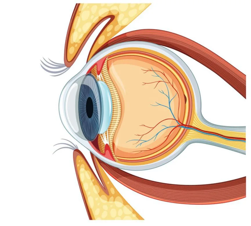
The eye, one of the most important sensory organs in the human body, has a complex structure that enables the process of vision. Eye anatomy is of great importance for protecting eye health and for diagnosing and treating eye related problems.
Eye anatomy consists of three main layers: the outer layer, the middle layer, and the inner layer.
- Outer Layer (Sclera and Cornea): The outer layer includes the sclera, a tough layer that protects the eye, and the cornea, which forms the clear front part. The cornea allows light to enter the eye and helps focus the image.
- Middle Layer (Choroid, Iris, Ciliary Body, and Cornea): The middle layer contains the blood vessels and nourishing tissues inside the eye. The choroid contains the vessels supplying the retina. The iris is the colored layer that determines eye color and controls the pupil. The ciliary muscle changes the shape of the eye's lens to enable focusing.
- Inner Layer (Retina): The inner layer contains the photoreceptor cells of the eye and converts light into nerve impulses that are sent to the optic nerve. This makes it possible for images to be transmitted to the brain.
Understanding eye anatomy is quite important for protecting eye health and detecting vision disorders. Components such as the cornea, iris, retina, and optic nerve play critical roles in how the eye functions. Eye doctors carefully examine these complex structures to assess eye health and diagnose vision disorders.
Regular eye exams are important for protecting eye health and for the early detection of vision disorders. These exams offer an effective way to examine eye anatomy and assess vision. For a healthy life, it is important not to skip regular eye checkups.
The Structure of the Eye: Main Parts and Their Functions
The eye is an important sensory organ with a complex structure that allows living organisms to detect light in their surroundings, form images, and relay them to the brain. Eye anatomy, together with the optical system and neural connections, works in perfect coordination to sense light and process visual information. The main parts of the eye and their functions are as follows:
1. Cornea: The Eye's Transparent Window
The cornea is a transparent, dome shaped structure that forms the outer layer of the eye. Positioned over other structures such as the iris and lens, this protective dome covering the front of the eye is called the cornea. The cornea makes an important contribution to protecting the inner parts of the eye by preventing potential damage from various environmental influences. Its layers help a smooth, healthy cornea correctly refract light and produce clear images.
2. Iris and Pupil: The Light Control Center
The iris is the ring shaped structure that forms the colored part of the eye. It determines eye color and contains the muscles that regulate the pupil. The pupil is the dark, round opening at the center of the iris. Its size changes depending on the amount of light. The iris and pupil control light by regulating how much enters the eye, producing a clear image.
3. Lens: The Key to Forming an Image
The lens is a transparent, elastic structure located between the iris and the retina. Its main purpose is to refract and focus incoming light rays to form a clear image on the retina.
The lens's flexibility allows the eye to focus on distant or near objects. This way, the image that is projected upside down on the retina is corrected and a clear image is formed.
Eye anatomy functions through a complex interaction between the cornea, iris, lens, and other structures. These structures ensure that the process of vision takes place in a healthy way. Regular eye exams are important for protecting eye health and for the early detection of vision disorders. For good vision health, it is important to understand how eye anatomy works and to pay attention to eye health.
Retina: The Point of Light Refraction
The retina is a critical structure located at the back of the eye and represents the important stage where light is converted into an image. At the start of the vision process, light detected at the back of the eye is perceived as an image on the retina. Light focused by the lens falls onto the photoreceptor cells of the retina. These photoreceptors convert light into electrical signals that are sent to the brain.
The retina contains two types of photoreceptor cells called cones and rods. Rods are effective in low light levels and night vision, but they cannot perceive color and have lower resolution. Cones provide higher resolution for detecting detail. The retina has special regions, such as the fovea, where cone cells are concentrated and the sharpest vision is achieved.
The layer located just beneath the retina that supports the photoreceptor cells is called the retinal pigment epithelium. This layer helps visual information be correctly perceived and processed.
The optic nerve is a bundle of nerve fibers that begins at the back of the eye and extends toward the brain. The nerve cells in the retina form the start of the optic nerve. These nerve cells gather information from the photoreceptor cells and carry it along the optic nerve to the brain. The optic nerve fibers cross behind the eyes, combining image information from both eyes to form the signals sent to the brain's visual centers. This complex structure of eye anatomy plays a critical role in relaying light to the brain to form an image.
Intraocular Pressure and Glaucoma
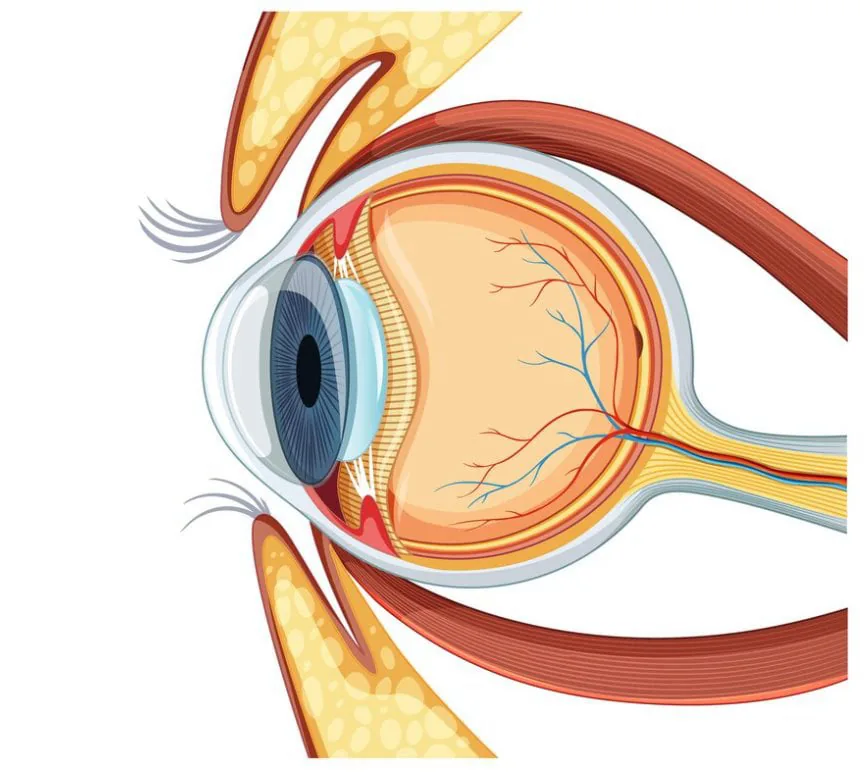
Special fluids in the front and back regions of the eye play an important role in regulating intraocular pressure and protecting eye health. One of these fluids, the "aqueous humor," is found in the front of the eye, while the other, the "vitreous humor," is found in the back of the eye. The balanced production and circulation of these fluids helps keep intraocular pressure under control. However, if this balance is disrupted, a serious eye disease such as glaucoma can develop.
Aqueous Humor: An Important Eye Fluid
Aqueous humor is a clear fluid found in the front part of the eye. Located in the region called the anterior chamber, this fluid plays an important role in maintaining the eye's shape and transparency. It is continuously produced and kept in balance through drainage channels. Aqueous humor helps regulate intraocular pressure while also supporting the eye's mechanical structure.
Vitreous Humor: Supporting Visual Information
Vitreous humor is a gel like fluid found in the back part of the eye, located in the region called the vitreous cavity. By supporting the shape of the retina, it helps images be perceived clearly. It also protects the eye's physical structure and helps keep the retina properly positioned. Intraocular pressure is kept under control through the balance and circulation of the aqueous and vitreous humor.
However, in some cases intraocular pressure can rise, which can lead to a serious eye disease called glaucoma. Glaucoma is typically characterized by elevated pressure resulting from conditions that block the drainage of aqueous humor. This can put pressure on the optic nerve and, over time, cause damage to the nerve fibers.
Regular eye exams are of great importance for maintaining the healthy functioning of eye anatomy and for detecting eye related problems at an early stage. Intraocular pressure and fluid balance are critical factors in protecting eye health.
As İdeal Göz Clinic in Kayseri, our goal is to provide you with the best possible eye care and to ensure your eyes are in ideal condition in every sense. With our warm and experienced staff, we will be by your side at every step, offering solutions tailored to you.
Call us now to schedule your eye exam and take action for your eye health! Remember, healthy eyes let you see a clearer world.
- We wish you healthy days,
- İdeal Göz Clinic, Kayseri
- Sources:
- https://www.britannica.com/science/human-eye
- https://www.aao.org/eye-health/anatomy/parts-of-eye
- https://www.ncbi.nlm.nih.gov/books/NBK11120/