














Cornea
Keratoconus
In keratoconus the cornea thins and bulges forward. Corneal crosslinking can stop it progressing.
Doctors in this field



What Is Keratoconus?
Keratoconus is the thinning and cone-shaped bulging of the cornea, the clear front layer of the eye. This bulging of the cornea causes irregular astigmatism and myopia, impairing vision. Patients with keratoconus experience symptoms such as blurred vision, double vision, glare, difficulty driving at night (particularly due to reflections from car headlights), and headache.
Its exact cause is not fully known, but genetic predisposition and mechanical factors such as eye rubbing play an important role in its development. Because allergic eye conditions, and therefore eye rubbing, are common in Turkey, keratoconus is seen more often here than in other countries.
Keratoconus usually begins during adolescence and progresses into a person's thirties. The disease begins with blurred vision. Because the vision disorder progresses rapidly, the patient changes eyeglasses frequently. After some time, patients begin to lose clear vision even with eyeglasses. A definitive diagnosis of keratoconus is made with corneal topography.
What Are the Treatments for Keratoconus?
Keratoconus treatment should be considered in 2 categories.
1. Treatments That Halt the Disease:
- Corneal crosslinking
2. Treatments That Improve Vision:
- Eyeglasses
- Soft contact lenses
- Rigid contact lenses (corneal rigid lenses, hybrid rigid lenses, scleral lenses)
- Topography-guided excimer laser (topo-laser)
- Corneal rings (intrastromal corneal ring segments)
Corneal Crosslinking
In keratoconus patients, this treatment aims to strengthen the weakened cornea and halt the disease. It is the only treatment that can stop the disease from progressing.
In crosslinking treatment, the surface cell layer of the cornea is first removed. Riboflavin drops are then applied for 30 minutes, followed by 30 minutes of ultraviolet A light exposure. A contact lens is placed at the end of the procedure and removed on day 5.
After crosslinking, the patient will have stinging, watery eyes, and light sensitivity for 2 days, along with mild blurred vision lasting about 1 month.
Eyeglasses
Patients with early-stage keratoconus can see well with eyeglasses. However, it must be remembered that, apart from crosslinking, no treatment, including eyeglasses, halts the disease. For this reason, one must always be certain that the disease has stopped progressing.
Soft Contact Lenses
Keratoconus patients who can see well with eyeglasses but do not want to wear them can use soft contact lenses. However, it must be remembered that mechanical trauma plays a role in the progression of keratoconus, and the soft lens itself, or the act of inserting and removing it, can cause such trauma.
Rigid Contact Lenses
In moderate and advanced keratoconus, the best quality vision is achieved with rigid contact lenses. Depending on the stage of keratoconus, a corneal rigid lens, hybrid lens, or scleral lens is prescribed. The biggest disadvantage of rigid lenses is that fitting them is difficult and adapting to them can be challenging. For this reason, the physician fitting the lens must be experienced in cornea and keratoconus care, and the eye center must have a full set of trial lenses available.
Topography-Guided Excimer Laser (Topo-Laser)
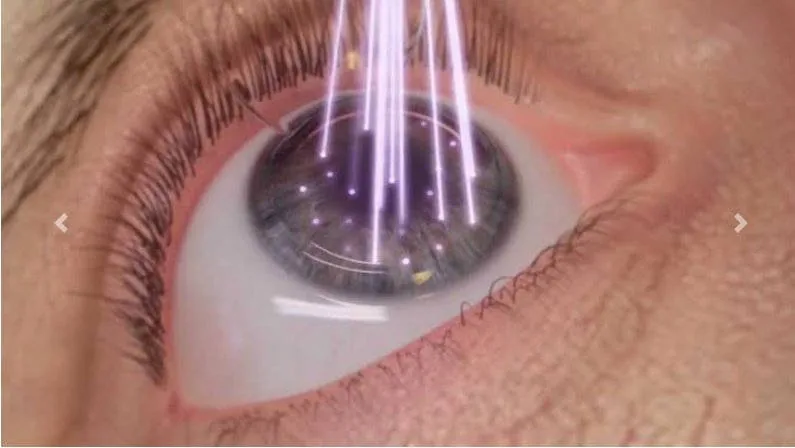
In the past, excimer laser (laser eye surgery) could not be performed on keratoconus patients. However, with the advent of crosslinking treatment, excimer laser can now be performed on keratoconus patients together with crosslinking. This excimer laser procedure must not be a standard one, however, because standard excimer laser is designed for a healthy cornea, whereas the cornea of a keratoconus patient is not normal. If excimer laser is to be performed on a keratoconus patient, it must always be topography-guided. This way, the laser not only lowers the diopter but also reduces the corneal irregularity caused by keratoconus, improving the quality of vision. This specialized treatment can only be performed with a physician experienced in keratoconus and an advanced excimer laser device.
Corneal Rings
Corneal rings are transparent segments implanted inside the cornea. The purpose of implanting corneal rings is to make the irregular cornea in keratoconus more regular, thereby reducing astigmatism. They are used in early and moderate stage keratoconus. Today, corneal rings are implanted using a laser, which has greatly reduced ring-related side effects. Their most significant disadvantage is glare at night, which some patients experience. The most important advantage of corneal ring treatment is that it is reversible.